One of the difficulties when trying to image biological tissue using optical techniques is that tissue scatters light, which makes it opaque. This scattering occurs because the different components of tissue, such as water and lipids, have different refractive indices, and it limits the depth at which light can penetrate.
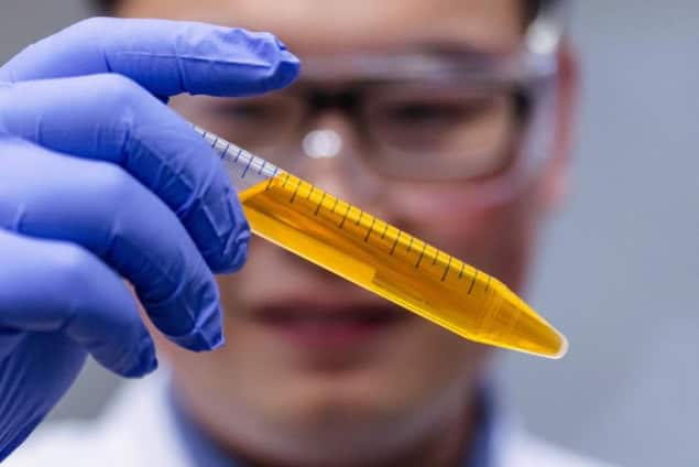
A team of researchers at Stanford University in the US has now found that a common water-soluble yellow dye (among several other dye molecules) that strongly absorbs near-ultraviolet and blue light can help make biological tissue transparent in just a few minutes, thus allowing light to penetrate more deeply. In tests on mice skin, muscle and connective tissue, the team used the technique to observe a wide range of deep-seated structures and biological activity.
In their work, the research team – led by Zihao Ou (now at The University of Texas at Dallas), Mark Brongersma and Guosong Hong – rubbed the common food dye tartrazine, which is yellow/red in colour, onto the abdomen, scalp and hindlimbs of live mice. By absorbing light in the blue part of the spectrum, the dye altered the refractive index of the water in the treated areas at red-light wavelengths, such that it more closely matched that of lipids in this part of the spectrum. This effectively reduced the refractive-index contrast between the water and the lipids and allowed the biological tissue to appear more transparent at this wavelength, albeit tinged with red.
In this way, the researchers were able to visualize internal organs, such as the liver, small intestine and bladder, through the skin without requiring any surgery. They were even able to observe fluorescent protein-labelled enteric neurons in the abdomen and monitor the movements of these nerve cells. This enabled them to generate maps showing different movement patterns in the gut during digestion. They were also able to visualize blood flow in the rodents’ brains and the fine structure of muscle sarcomere fibres in their hind limbs.
Reversible effect
The skin becomes transparent in just a few minutes and the effect can be reversed by simply rinsing off the dye.
So far, this “optical clearing” study has only been conducted on animals. But if extended to humans, it could offer a variety of benefits in biology, diagnostics and even cosmetics, says Hong. Indeed, the technique could help make some types of invasive biopsies a thing of the past.
“For example, doctors might be able to diagnose deep-seated tumours by simply examining a person’s tissue without the need for invasive surgical removal. It could potentially make blood draws less painful by helping phlebotomists easily locate veins under the skin and could also enhance procedures like laser tattoo removal by allowing more precise targeting of the pigment beneath the skin,” Hong explains. “If we could just look at what’s going on under the skin instead of cutting into it, or using radiation to get a less than clear look, we could change the way we see the human body.”
Hong tells Physics World that the collaboration originated from a casual conversation he had with Brongersma, at a café on Stanford’s campus during the summer of 2021. “Mark’s lab specializes in nanophotonics while my lab focuses on new strategies for enhancing deep-tissue imaging of neural activity and light delivery for optogenetics. At the time, one of my graduate students, Nick Rommelfanger (third author of the current paper), was working on applying the ‘Kramers-Kronig’ relations to investigate microwave–brain interactions. Meanwhile, my postdoc Zihao Ou (first author of this paper) had been systematically screening a variety of dye molecules to understand their interactions with light.”

Mouse brain imaging reaches record-breaking resolution
Tartrazine emerged as the leading candidate, says Hong. “This dye showed intense absorption in the near-ultraviolet/blue spectrum (and thus strong enhancement of refractive index in the red spectrum), minimal absorption beyond 600 nm, high water solubility and excellent biocompatibility, as it is an FD&C-approved food dye.”
“We realized that the Kramers-Kronig relations could be applied to the resonance absorption of dye molecules, which led me to ask Mark about the feasibility of matching the refractive index in biological tissues, with the aim of reducing light scattering,” Hong explains. “Over the past three years, both our labs have had numerous productive discussions, with exciting results far exceeding our initial expectations.”
The researchers say they are now focusing on identifying other dye molecules with greater efficiency in achieving tissue transparency. “Additionally, we are exploring methods for cells to express intensely absorbing molecules endogenously, enabling genetically encoded tissue transparency in live animals,” reveals Hong.
The study is detailed in Science.


