Using a scaffold made from conductive silicon nanowires, researchers in the US have developed artificial heart tissue that they say could be readily transplanted into natural tissue. Led by Ying Mei at Clemson University, the team hopes its technique could be a game changer in the global effort to treat heart disease.
Together, cardiovascular diseases are the leading cause of death worldwide, taking an estimated 17.9 million lives every year according to the World Health Organization. One of the main reasons these conditions are so prevalent is that heart cells have a limited ability to regenerate themselves when damaged, making it especially challenging for researchers to develop effective treatments.
Among the most promising advances in heart disease research involves the use of heart cells derived from stem cells, which are usually injected straight into damaged heart muscle. So far, this technique has been used to restore heart contraction in several different types of animal – but is still a long way from becoming a medically viable treatment. Among the things holding the technique back include the low survival rate of injected cells, and a limited recovery of the heart’s full function – especially the regular rhythm of its contraction.
Miniaturized, organ-like structures
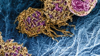
Recently, progress in stem cell treatments has been achieved for a variety of other organs, including the brain, lung and retina. Each of these studies involved the transplantation of organoids. These are miniaturized, organ-like structures that can be grown in the lab from stem cells, and which replicate the structure and function of a real organ.
Although cardiac organoids have already proven to be an excellent platform for modelling heart disease and testing new drugs, their potential for treating heart disease still requires further investigation.
In their study, Mei’s team investigated whether cardiac organoids could be made to contract in regular rhythms by growing the tissue in scaffolds made from electrically conducting silicon nanowires.
Biocompatible and biodegradable
In biological applications, silicon offers a host of advantages compared with other conducting nanomaterials. Through a series of tests on heart tissue in rats, the team showed that the material is biocompatible, biodegradable, has an easily tuneable conductivity, and easily adjustable dimensions and surfaces – all of which would be vital to ensuring the greatest chance of success for an implanted organoid.
Through a carefully controlled process, Mei and colleagues created an organoid from a mixture of stem-cell-derived heart cells, stromal connective tissue cells, and endothelial cells – which line the walls of blood vessels.

Ultrathin e-tattoo provides continuous heart monitoring
In the experiment, these cells assembled themselves around a pre-constructed silicon scaffold to form a nanowired organoid. Just as the team hoped, this miniature tissue performed many of the heart’s most important functions, including its regular rhythm of contraction.
When the researchers injected their nanowired organoids into rat hearts, they recorded a far higher cell survival rate compared with unwired organoids. This accelerated the development of its stem cells into healthy, well-functioning heart tissue.
Mei’s team hopes its research could be an important milestone towards feasible new treatments for heart disease. If the same success can be recreated with organoids grown from human stem cells, it could pave the way for treatments which enable patients’ heart tissue to regenerate and restore its full function. In turn, the use of silicon nanowire scaffolds may ultimately lead to new treatments tailored to different types of heart disease, with the potential to save millions of lives.
The research is described in Science Advances.


