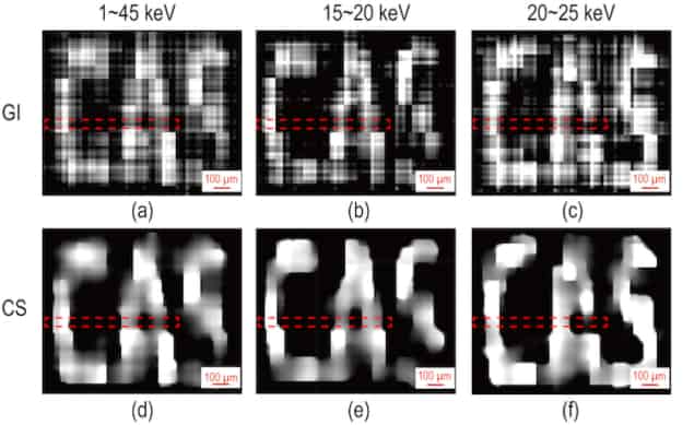
Researchers in China have used an imaging approach known as X-ray ghost imaging (XGI) to obtain spectral images of an object using a single-pixel detector. This technique could find applications in a host of different fields, including biological and medical imaging, materials science and environmental sensing.
Unlike conventional cameras, ghost imaging does not directly capture an image of an object. Instead, it reconstructs the image from the correlations between the light that the object reflects or transmits and a series of “speckle patterns” used to illuminate it. In classical ghost imaging, these patterns are produced by two beams: one that encodes a random pattern that acts as a reference, but which doesn’t directly probe the sample, and another that passes through the sample. The two beams both acquire partial information about the object, but neither one alone can form a complete image.
Modulating mask
In their work, researchers led by Ling-An Wu and Li-Ming Chen of the Institute of Physics, Chinese Academy of Sciences, Beijing, began by modulating an X-ray beam using the patterns in a 2-inch square mask made of gold and placed about 45 cm from the X-ray source. They then passed the resulting structured beam through their sample, which was located about 1 cm away from the mask. Since the sample is so close to the mask, the pattern of X-rays projected through the sample is the same as the pattern in the mask itself – a prerequisite for ghost imaging in this work. This orientation also ensures that the resolution is practically the same as the 10 μm pixel size of the mask.
To block unwanted radiation, the researchers inserted a 3-mm-thick square aperture made of copper between the object and the detector. To collect their data, they placed a “bucket” spectrometer (comprising a sensor, signal amplifier, digital pulse processor and specialized software) 25 cm behind this aperture.
Wide range of different X-ray energies
The 5 × 5 mm2 sensor efficiently and simultaneously detects a wide range of X-ray energies (from 3 to 45 keV) as they are transmitted through the sample with a resolution of 1.5 keV, Wu and colleagues say. The key component of this single-pixel detector is a 1-mm-thick cadmium telluride diode that produces current pulses proportional to the energy of the incident X-ray photons. These analogue intensity signals are then converted to digital pulses of different heights. Next, the output signals are reshaped and amplified, before being processed to produce an energy spectrum.
After recording all this spectral information, the researchers built up X-ray ghost images of the sample by measuring the correlations between different intensities of light transmitted by the object through the mask. This is known as second-order correlation, and is a routine technique in ghost imaging.
The technique is the first demonstration of energy-selective X-ray ghost imaging, where spectral and intensity information can be obtained at the same time without affecting each other, Wu says. The X-ray source the Beijing group employed is also simpler than the synchrotron radiation sources used in previous studies. “Our source is a conventional table-top X-ray tube that emits polychromatic rays of a much lower intensity,” she explains. “The key component in our experiments is, in fact, our specially-fabricated etched gold modulation mask. With modern micromanufacturing techniques, we can etch most of the patterns in the mask as designed. We characterized ours using scanning electron microscopy, from which we confirmed that the pixel size was indeed 10 μm.”

Terahertz microscope produces highly accurate ‘ghost’ images
Attractive for medical imaging
Since X-ray ghost imaging can be performed with much lower levels of radiation than traditional X-ray imaging, it should be attractive for medical imaging and analysing in vivo samples, she tells Physics World. “The technique acquires images according to the spectral fingerprint of the object, so different tissue layers can be distinguished more easily and without damage.”
Other application areas include analysing mineral ores and archaeological samples, she adds.
The Beijing team’s members, who report their work in Chinese Physics Letters (which is published jointly by IOP Publishing and the Chinese Physical Society), say they now plan to analyse real biological samples and other materials. “Analysing the X-ray signals from stellar objects is also another possibility,” Wu adds.


