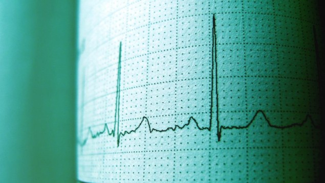
A simple set of mathematical rules that accurately identifies signs of competing electrical signals in the heart has been developed by Leon Glass at McGill University and colleagues. The Canadian team did in vitro experiments that showed how abnormal beating triggered by anomalous electrical impulses can generate distinctive triplets. These triplets also appear in cardiac data from real patients and emerge from a simple mathematical model.
Our heartbeat is controlled by a cluster of specialized cells called a pacemaker. Located in the right atrium, the pacemaker broadcasts regular electrical “sinus” signals that cause heart muscle cells to contract in a regular manner. However, the heart can also contain an abnormal “ectopic” pacemaker located in a different part of the organ. This pacemaker can send out electrical signals, often at lower frequencies than the sinus pacemaker. The heart muscles can respond to both sinus and ectopic signals, leading to an irregular beating of the heart called ectopic arrhythmia.
To monitor for signs of ectopic pacemakers, scientists have developed mathematical models that predict the patterns of irregular heart activity that can emerge from this interference. In this new study, Glass’s team did experiments that put these models to the test.
Genetically modified
The team began by creating artificial pacemakers in cardiac cells taken from mice. These cells had been genetically modified so that the voltages produced by their cell membranes can be controlled using light. In thin layers of heart cells, the researchers used short light pulses to create pairs of pacemaker signals with slightly different frequencies (one sinus and the other ectopic), at spots positioned 8.5 mm apart.
In the region between the spots, they measured the number of sinus pulses detected between successive ectopic pulses – where the ectopic pulses occur at a lower frequency than the sinus pulses. Interference between the pulses causes this number to vary according to when and where in the sample the measurements are made.

3D virtual heart helps pinpoint irregular heartbeat
Glass and colleagues discovered that the number of these intervening sinus pulses always came in distinctive triplets: such as 1, 2, 4; 1, 4, 6; or 4, 6, 11. These triplets all seemed to follow just two simple rules: that one number must be odd, and that the largest number must be one larger than the sum of the two smaller numbers.
Based on these rules, the researchers reproduced the same patterns in simple mathematical models, and also searched for the patterns in real electrocardiogram data from 47 patients. Despite the immense complexity of the heart, they detected the distinctive triplets in eight electrocardiograms.
Glass’s team hopes that the remarkable simplicity of the triplet pattern should make it easily detectable using wearable heartbeat monitors. This could strengthen the ability of cardiologists to diagnose and treat potentially life-threatening irregular heartbeats.
The research is described in Physical Review Letters.


