Chronic inflammation of the brain is linked to a range of increasingly common degenerative brain diseases, such as Alzheimer’s and Parkinson’s. Evidence suggests that neuroinflammation contributes to the progression and worsening of such diseases.
However, the current diagnostic tool used for monitoring brain inflammation, positron emission tomography (PET), involves ionizing radiation, which may increase the patient’s risk of developing cancer. The delivered radiation dose also makes it impractical to perform longitudinal research studies or repeat testing during treatments. As such, there’s a need to develop an efficient imaging modality that will not worsen the condition of patients with neuroinflammation.
Researchers from Alicante, Spain, have developed a non-invasive method for visualizing brain inflammation using diffusion-weighted magnetic resonance imaging (DW-MRI). The team, led by Silvia De Santis and Santiago Canals from the Institute of Neurosciences, a joint centre of the Spanish Superior Research Council (CSIC) and the Miguel Hernández University (UMH), devised a series of MR data acquisition sequences and mathematical models to detect changes in the activation of two brain cell types associated with neural inflammation: astrocytes and microglia.

DW-MRI enables the collection of images from microstructures within the brain with a high resolution by utilizing the random motion of water molecules within brain tissue. Most previous research use of DW-MRI has focussed on the brain’s white matter and axons, but to investigate chronic inflammation, the researchers were interested in imaging the brain’s grey matter.
To focus on the all-important astrocytes and microglia, they therefore had to adapt and design advanced DW-MRI sequences, for use in combination with mathematical models based on the biological knowledge of the functional tissues within the brain.
The scientists tested their model on rats, using an established technique for inducing inflammation (administering lipopolysaccharide), which first activates the microglia, followed by a delayed response from astrocyte cells, allowing independent investigation of the two cell types. The MRI scans showed specificity to both the microglial and astrocyte activation in grey matter in vivo.
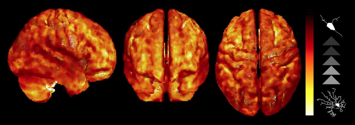
Secondly, the researchers used the method in human participants in a proof-of-concept experiment, scanning six healthy volunteers across five occasions. They found that the pattern of microglial cell density significantly correlated with the MRI parameter of stick fraction. The results highlight the model’s ability to detect glial biomarkers and confirmed reproducibility between scanning sessions.

Lesion mapping tool makes early signs of dementia easier to spot
The researchers hope that their work will enable better characterization of brain tissue microstructure during inflammation, with higher resolution and the possibility of longitudinal study without a dose of ionizing radiation. They believe that this could transform the diagnosis and treatment monitoring of the many diseases associated with an inflammatory glial response.
The research is published in Science Advances.


