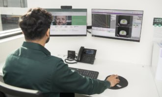
Using time-resolved CT data to estimate the breathing capacity in different parts of the lung could enable more effective treatment of lung tumours while reducing the risk of radiation-induced toxicity

Precision is everything in radiotherapy treatments, with the overriding objective to deliver the radiation dose directly to the tumour without causing any damage to healthy tissue and the surrounding organs. When treating the lung, for example, exposing the parts that are responsible for gas exchange to ionizing radiation can trigger a range of respiratory issues, ranging from short-term inflammation and shortness of breath to chronic conditions that can cause permanent scarring of the lung tissue.
“The lung is an important organ-at-risk, and radiation-induced toxicity is our main concern when treating patients with lung tumours,” says Florian Putz, a senior radiation oncologist at the University Hospital Erlangen in Germany. “The standard clinical practice is to minimize the dose across the entire organ, but that doesn’t take account of any spatial variation in the ventilation provided by different parts of the lung.”
Putz points out that the most functional parts of the lung – the areas that efficiently extract oxygen from the air we breathe – are not always equally distributed. Localized tissue scarring can compromise the gas-exchange process in some places, while any inflammation can constrict the airways in particular areas of the lung. Taking account of these spatial variations would enable clinicians to spare the parts of the lung that contribute most to a patient’s breathing capacity, and offers the potential to deliver more dose to areas where ventilation is already suppressed.
Putz has been investigating whether such spatial information on pulmonary function could be retrieved from the data recorded in time-resolved CT scans. Such 4DCT scans are routinely acquired during thoracic treatments, primarily to record the movement of the tumour as the patient breathes in and out, and a novel tool built into the syngo.via RT Image Suite from Siemens Healthineers uses this data to calculate and image the ventilation in the five distinct lobes of the lung. “This lung ventilation tool would enable us to spare the lobes that contribute most to the patient’s breathing capacity, and offers the potential of escalating the dose that is delivered to the lobes that are not so functionally relevant,” explains Putz.
Inside the tool is an automated deep-learning technique that uses the 4DCT images to segment the five lobes of the lung – first when the patient has taken a full inhalation and then when all the air has been expelled. The ventilation in each lobe is then calculated from the difference in air volume between those two states, and then expressed as percentage value by normalizing with the air volume at maximum exhalation.
“The segmentation gives us the total volume of the lung in each state, and then we calculate the air volume from a simple model that treats the lung as a linear combination of air and water,” explains Christian Möhler, product manager for radiotherapy imaging software at Siemens Healthineers. “This straightforward mathematical approach is robust to uncertainties, and we can use the percentage values to provide a colour-coded view of the ventilation in each of the five lobes.”
Other methods have been proposed to extract ventilation information from time-resolved CT, in many cases providing a granular view down to the level of individual voxels. In this case, however, each data point in the CT image must be correlated between the inspiration and expiration states, which can introduce inaccuracies when trying to map the intricate airways and blood vessels within the lung. In contrast, the algorithm developed by Siemens Healthineers avoids the need for this complex registration process, instead relying only on the segmentation of the five lobes that are produced for each breathing state.
“We need to avoid any errors that could lead to wrong treatment decisions, which means that the algorithm needs to be robust,” comments Putz. “The lobes also tend to be quite homogeneous, which means that the ventilation across the spatial compartment of each one tends to be quite similar.”
Putz has been working with medical physicist Juliane Szkitsak to compare the information extracted from the CT lung ventilation tool with the data from standard breathing tests that exploit spirometry to assess the overall pulmonary function of both lungs. From the ventilation metric provided by the software tool he calculated the volume of air across all five lobes of the lung, allowing a direct comparison with the results from these spirometry tests. “There was a high correlation between the two, which suggests that the tool is working as it should,” he says.
In a further evaluation at University Hospital Erlangen, Szkitsak replanned the treatment of six patients using information from the CT lung ventilation tool. “The new treatment plans avoid the healthy parts of the lung and only treat through the lobes that the ventilation data show might already be impaired,” she explains. “The additional information from the ventilation tool made the optimization more complex, but I think it would become quicker and easier as you get more familiar with it.”
However, Szkitsak also points out that for this small selection of patients it was difficult to assess the benefits of using the tool, since the complexity of thoracic treatment plans and the need to avoid other organs-at-risk makes it difficult to produce comparable plans. She also found that in some cases the new treatment plans resulted in the radiation beam passing through more of the lung, which was a cause for concern among the clinical team at Erlangen. “Usually we avoid as much lung volume as possible by, for example, minimizing the distance between the skin and the tumour,” she explains. “With the plans produced using the ventilation algorithm we sometimes had to go through the lung in a different way, with the result that more of the lung was exposed to radiation.”
With no clear evidence to show that the ventilation data can reduce the risk of damaging the lung, the clinicians at Erlangen have concluded that it is too early to change their treatment strategy. “While there is a growing consensus in the literature that CT lung ventilation produces valid results, all the physicians at our hospital need to be convinced that it can provide a benefit for patients without putting any of them at risk,” says Putz. “Additional studies and larger evaluations will be needed, while widespread acceptance will most likely require clinical trials that show reduced pulmonary toxicity in treatments that exploit such a tool.”
While the ventilation information might not yet influence the initial treatment planning process, Putz believes that it could help clinicians to monitor lung function during multiple rounds of treatment. Such regular monitoring could augment the standard breathing tests that are usually performed before treatment starts, helping clinicians to detect early signs of toxicity and even to adapt their treatment plans to take account of any changes in ventilation.
“We are moving towards adaptive radiotherapy, in which we take multiple scans as the treatment progresses and then update the treatment plan to reflect any changes in the body,” explains Putz. “Using this tool we have seen changes in the spatial distribution of the ventilation during treatment, for example in patients where pulmonary effusion caused fluid to accumulate in the lung, and having this information would enable us to update our treatment plan and deliver better outcomes for the patient.”


