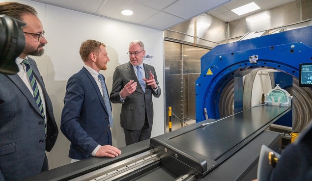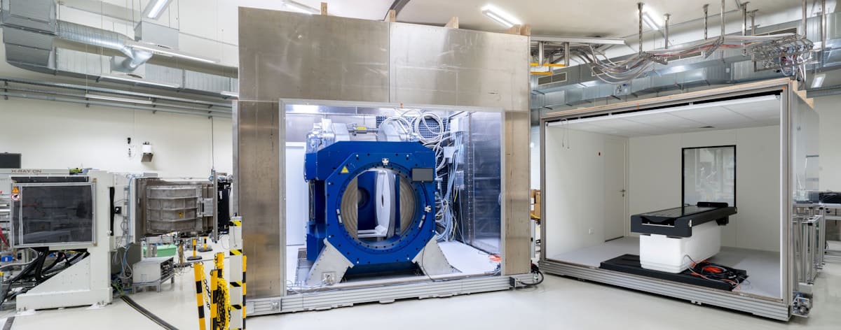
This week saw the official inauguration of the world’s first research prototype for whole-body MRI-guided proton therapy. The launch ceremony, at OncoRay – the National Center for Radiation Research in Oncology in Dresden, marked the start of scientific operation using the prototype, which is designed to enable real-time MRI tracking of moving tumours during proton therapy.
Proton therapy provides a means to treat tumours with extreme precision. The finite range of a proton beam enables extremely conformal dose targeting with reduced dose to nearby healthy tissue. This high conformality, however, makes proton treatments particularly sensitive to anatomical changes in the beam path, which can impair the targeting precision when treating a moving target. Real-time imaging during treatment could help solve this drawback by synchronizing dose delivery with the tumour position.
MRI provides a way to visualize moving tumours and surrounding healthy tissue with excellent soft-tissue contrast. Performing MRI during treatment delivery could enable real-time visualization of tumour motion and the potential for real-time adaptation. MRI can also detect any anatomical changes between consecutive treatment sessions.
And while MRI guidance for photon-based radiotherapy is now commercially available and in clinical use, OncoRay’s is the first such system that exploits MRI to guide proton therapy.
The prototype device – developed by OncoRay’s Experimental MR-Integrated Proton Therapy research group, led by Aswin Hoffmann – combines a horizontal proton therapy beamline with a whole-body MRI scanner that rotates around the patient. Hoffmann notes that this geometry enables innovative patient positioning approaches, including treatment in both lying or in upright positions.

The ultimate goal of the OncoRay team, along with scientists from the Helmholtz-Zentrum Dresden-Rossendorf (HZDR) and the Dresden University Medical Center, is to use real-time MRI to monitor cancer patients during their treatments and significantly improve the targeting accuracy of proton therapy.
Hoffmann tells Physics World that the first study using the new prototype MRI–proton therapy system is designed to assess the mutual electromagnetic interactions between the proton pencil-beam scanning (PBS) beamline and the in-beam MRI scanner. “We need to answer two questions,” he explains, “do the magnetic fringe fields produced by the PBS beamline affect the MR image quality during irradiation and does the static magnetic field of the MRI system affect the beam delivery system?”

One step closer to real-time MR imaging in proton therapy
In the longer term, the researchers will use the prototype to demonstrate the added value that whole-body, real-time MRI guidance could bring to treatments of mobile tumours in the chest, abdomen and pelvis.
“I have been working on this specific project since 2018, the last three years of which [I worked] very intensively with the industry partners involved. I am proud that together we have managed to realize and put this system into operation. I look forward with great anticipation to the next phase in which the scientific challenges will be addressed,” says Hoffmann.


